Understanding the Impact of Chronic Back Pain: Sara Guilcher in the New York Times
Our colleague Dr. Sara Guilcher was recently interviewed in a significant article examining chronic back pain in society, prompted by recent events in New York involving the CEO UnitedHealthcare. The circumstances are tragic. The coverage has sparked an important conversation about chronic pain and its far-reaching effects.
The article highlights that approximately 16 million adults in the United States experience persistent back pain that interferes with daily activities, making it one of the most common medical challenges and the leading cause of disability worldwide according to the WHO. Similarly, chronic pain affects 1 in 5
The piece emphasizes how chronic back pain can fundamentally affect every aspect of a person's life - from emotional wellbeing and sleep to work and social relationships. It features insights from multiple medical experts, including Dr. Guilcher, discussing the often-overlooked psychological and social impacts of chronic pain conditions.

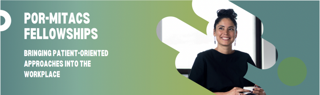
POR-Mitacs Fellowship
PASSERELLE, SPOR National Training Entity, and Mitacs invite undergraduate, college and graduate students, and postdoctoral fellows from all disciplines to participate in this 2nd edition of the PatientOriented Research (POR)-Mitacs Fellowship Program.
PASSERELLE, SPOR National Training Entity aims to collectively grow, support, and sustain the capacity for a collaborative, interdisciplinary, and innovative patient-oriented research (POR) environment capable of addressing evolving healthcare questions and contributing to enhancing patients’ healthcare experience and well-being. In support of this goal, a POR-Mitacs Fellowship Program was established to provide a mechanism to support projects that integrate the principles of POR and provide fellows with training, networking and mentoring opportunities that foster the development of POR and professional skills that are applicable across diverse career trajectories.
If you are interested in applying, contact your local Mitacs Advisor and PASSERELLE at info@passerelle-nte.ca to learn more or discuss the next steps. The Mitacs Advisor will be able to clarify the application process and requirements. Please see here for more information.

Job Posting - Post Doctoral Researcher - UHN
Dr. Andrea Furlan is hiring a Postdoctoral Researcher to start in January 2025. The successful candidate will work with a multidisciplinary team of clinicians and educators to develop and update materials for guideline implementation. The candidate will gain skills in implementation science, continuing medical education, and the appropriate use of opioids for chronic non-cancer pain. Please see the Job ad here.


#NPAW November 3-9
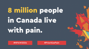
National Pain Awareness week: November 3-9, 2024
Dear Members,
National Pain Awareness Week (NPAW) is just around the corner, from November 3 - 9, 2024. The University of Toronto Centre for the Study of Pain (UTCSP) and its members are leaders in pain research, education, and knowledge translation, and are championing many of the NPAW events. You can get involved too, by helping to spread the word, attending the UTCSP NPAW talk, and exploring a variety of in-person and virtual events focused on advancing the understanding of pain and the pain experience.

Collaborating on Pain
By Diane Peters
Pain Week and other ways the U of T Centre for the Study of Pain tackles this complex experience
National Pain Awareness Week is coming up on November 3 to 9. The best place to discover more about the complexities of pain during this week — and beyond — is the University of Toronto Centre for the Study of Pain (UTCSP).
At this virtual centre, the Faculty of Dentistry takes the lead, working with the faculties of Medicine, Pharmacy and Nursing to tackle pain.

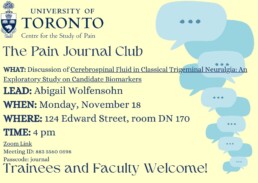
UTCSP Presents The Pain Journal Club: November 18
Attention Trainees and Faculty!
We are pleased to share an exciting opportunity for learning and networking: The Pain Journal Club. Happening on most Mondays throughout this term and next, join others in the sphere of pain lead discussions on timely and relevant research in the field.
LEAD: Abigail Wolfensohn
WHEN: Monday, November 18
WHERE: 124 Edward Street, room DN 170
TIME: 4 pm
Meeting ID: 883 5560 0598
Passcode: journal

Laura Murphy's Career
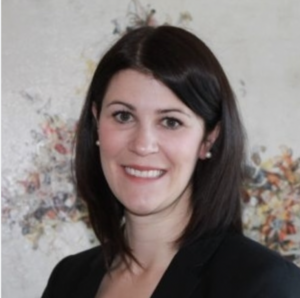
Laura graduated from the Leslie Dan Faculty of Pharmacy, University of Toronto with her Bachelor of Science in Pharmacy in 2006. Following graduation, Laura completed her hospital residency (Hospital for Sick Children, 2007) and she continued her academic pursuits at the University of Toronto by earning her Doctor of Pharmacy degree in 2009. Laura’s passion for learning continued throughout her career with the more recent successful completion of her Masters of Science in Pharmacy from the University of Toronto in 2020. During her undergraduate studies, she worked as a pharmacy student with the Ministry of Health and Long-Term Care, a research assistant with the Pharmacy Post publication, as well as in Drug Information at St Michael’s Hospital. After the completion of her residency and postbaccalaureate studies, Laura started as a Clinical Pharmacist Practitioner at North York General Hospital in 2009. She joined University Health Network in the fall of 2010, first at the Toronto Western Hospital (Altum Health Clinic), then later went on to accept a role as interim Pharmacy Clinical Site Leader (CSL) at Toronto General Hospital, and then transitioned to the role of CSL at Toronto Rehabilitation Institution in May 2012.
Laura’s love of research was sparked early in her career. Laura went on to become a Clinician Investigator with the KITE Research Institute. Her academic work focused on pain management, including multiple grants to understand the relationship between cannabis and opioid use (CIHR Operating Grant), examining the relationship amongst opioid subjective effects and pharmacokinetics of extended release opioids at shortened dosing intervals in patients with chronic pain (CSHP Research and Education Grant), and more recently, looking at establishing the role of community pharmacists in providing care for people using cannabis (Canadian Foundation for Pharmacy). She also co-led the development of a competency-based program for hospitals pharmacists (CSHP Research and Education Grant). Laura collaborated broadly with interprofessional researchers and clinicians in medicine and pharmacy and had a prolific track record of peer-reviewed journal publications, journal articles, conference abstracts and magazine entries. She was first author on many peer-reviewed papers on the topic of pain management in journals such as The Canadian Journal of Pain, Canadian Medical Association Journal (CMAJ), Canadian Journal of Hospital Pharmacists, International Journal of Clinical Pharmacy, and the Journal of Pain Research, and was co-author on many others.
Laura was regularly sought out for her expertise in pain therapeutics, including as an Executive Committee Member and eventually Co-chair for the University of Toronto Centre for the Study of Pain Interfaculty Pain Curriculum, and an Expert Advisory Committee Member to the Canadian Pain Task Force. She was also soughtafter as a participant in local, provincial and national meetings, as an invited speaker at the Ontario Medical Association Physician Health Program, Ontario HIV Treatment Network, Ontario Pharmacists Association Conferences, Canadian Pharmacists Conference, and the Canadian Society of Hospital Pharmacists Professional Practice Conference. In addition, she served as a consultant to the Canadian Agency for Drugs and Technologies in Health, as well as the OpiATE project focusing on pharmacist mentoring in Addiction and Pain (CAMH). Laura was an accomplished educator and had many diverse teaching credits throughout her career. She held numerous teaching roles at the University of Toronto, including teaching assistant, evaluator and course coordinator with the undergraduate Pharmacy program, preceptor with the Doctor of Pharmacy program and Entry Level Doctor of Pharmacy program, instructor with the PharmD for Pharmacists program, teaching assistant for the Physician’s Assistant Program, facilitator for the Interfaculty Pain Curriculum, and instructor in the Faculty of Medicine, She was an expert lecturer on pain for the undergraduate and Doctor of Pharmacy programs, Faculty of Nursing and the Connaught Pain Institute. In addition, she served as a teaching assistant in critical appraisal, rehabilitation and pain at the University of Alberta.
Laura was the distinguished recipient of many awards at the local, provincial and national levels. Some notable examples include the Martin Wolfish MD Awards for excellence in Clinical Teaching (2011), the Pharmacy Professor’s Awards from the Leslie Dan Faculty of Pharmacy (2015), and the Management and Leadership Best Practices Award from the Canadian Society of Hospital Pharmacists in 2017. In 2018, she was the recipient of the Clinical Research Student Seminar Group Poster Awards at the Graduate Research in Progress Symposium (University of Toronto), and was awarded the Ontario Branch, CSHP E. Amy Eck Award for Hospital/Outpatient Pharmacy-based project as well as the Literary Award focused on a Therapeutic Review. Throughout her career, Laura had a profound impact on all those around her. She was truly committed to helping patients with chronic pain manage their medications as well as teaching other health professionals to develop their own understanding and comfort in pain management. For those of us that have had the pleasure of working with Laura, we will remember her as a generous, selfless and compassionate leader, who cared deeply about her team. She thrived on seeing her staff grow as practitioners and researchers themselves. We will always remember the gentle way in which Laura pushed each of us to be the best version of ourselves. Our thoughts are with Laura’s family, friends, colleagues and all those whose lives and careers she touched in big and small ways.
Summary prepared by Jennifer Harrison & Aleesa Carter
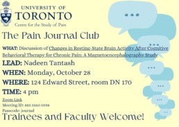
UTCSP Presents The Pain Journal Club: October 28
Attention Trainees and Faculty!
We are pleased to share an exciting opportunity for learning and networking: The Pain Journal Club. Happening on most Mondays throughout this term and next, join others in the sphere of pain lead discussions on timely and relevant research in the field.
WHAT:Discussion of Changes in Resting-State Brain Activity After Cognitive Behavioral Therapy for Chronic Pain: A Magnetoencephalography Study
LEAD: Nadeen Tantash
WHEN: Monday, October 28
WHERE: 124 Edward Street, room DN 170
TIME: 4 pm
Meeting ID: 883 5560 0598
Passcode: journal
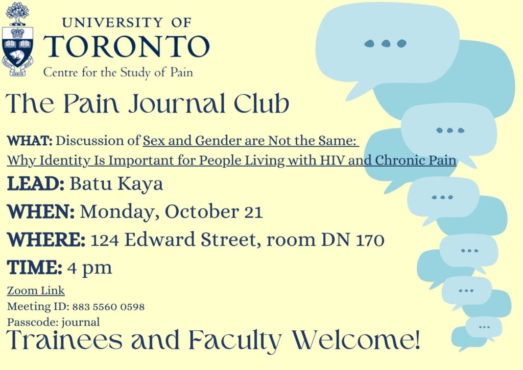
UTCSP Presents the Pain Journal Club - October 21, 2024
Attention Trainees and Faculty!
We are pleased to share an exciting opportunity for learning and networking: The Pain Journal Club. Happening on most Mondays throughout this term and next, join others in the sphere of pain lead discussions on timely and relevant research in the field.
WHAT: Discussion of Sex and Gender are Not the Same: Why Identity Is Important for People Living with HIV and Chronic Pain
LEAD: Batu Kaya
WHEN: Monday, October 21
WHERE: 124 Edward Street, room DN 170
TIME: 4 pm
Meeting ID: 883 5560 0598
Passcode: journal